Blank Diagram Of A Long Bone : BIO 430 Study Guide (2012-13 Smith) - Instructor Smith at ... : Each end is filled with a lattice.. Learn long bone diagram with free interactive flashcards. A long bone is a bone that has a shaft and 2 ends and is longer than it is wide. Long bones are one of the five bone types that are classified by shape. The bundle gives you access to a second set of diagrams, studying the anatomy of a long bone. The structure of a long bone allows for the best visualization of all of the parts of a bone (figure 1).
The blood vessels inside a bone. The tough membrane covering the shaft of the bone. The tarsus or heel bone consist of 7 bones that make up the posterior part of the foot, that is present between the tibia, fibula and metatarsals. Long bones — a subtype of bones — are longer than they are wide. Smartdraw includes 1000s of professional healthcare and anatomy chart templates that you can modify and make your own.
Skeletal System - Catherine Dela Cruz from caathdelacruz.weebly.com The ends of a long bone contain spongy bone and an epiphyseal line. The structure of a long bone allows for the best visualization of all of the parts of a bone (figure 1). The blood vessels inside a bone. Inside this is a layer of spongy (cancellous) bone which contains red bone marrow. Cheek bone (zygoma) upper jaw (maxilla). Short bones provide stability and support as well as. The end of a long bone. Labelled diagram of long bone.
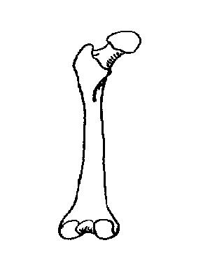
Image of a typical long bone is shown with numbers identifying the various parts, such as the epiphysis.
The femur and/or hip may fracture secondary to trauma, so understanding the femur bone anatomy is important. It is located between the elbow joint and the shoulder. At the elbow, it connects primarily to the ulna, as the forearm's radial bone connects to the. Long bones include the humerus (upper arm), radius (forearm), ulna (forearm), femur (thigh), fibula (thin bone of the lower leg), tibia (shin bone) , phalanges (digital bones in the hands and feet), metacarpals (long bones within the hand), and metatarsals (long bones. The tarsus or heel bone consist of 7 bones that make up the posterior part of the foot, that is present between the tibia, fibula and metatarsals. The long bones of the body contain many distinct regions due to blank bone diagram rome fontanacountryinn com. This is an online quiz called long bone diagram. Image of a typical long bone is shown with numbers identifying the various parts, such as the epiphysis. After publishing this diagram of a long bone we can guarantee to aspire you. The tough membrane covering the shaft of the bone. The diaphysis is the tubular shaft that runs between the proximal and distal ends of the bone. The humerus is the long bone in the upper arm. Blank bone diagram rome fontanacountryinn com.
Skeletal system test bank questions contain over 100 questions you can customize for your students, including multiple choice, true and false, labeling, fill in the blank, matching, short answer and long answer questions. A long bone has two parts: Long bones have a thick outside layer of compact bone and an inner medullary cavity containing bone marrow. Label the parts of a long bone. A long bone is a bone that has a shaft and 2 ends and is longer than it is wide.
Clavicle Bone at Long Beach City College - StudyBlue from classconnection.s3.amazonaws.com There is a printable worksheet available for download here so you can take the quiz with pen and paper. Most, but not all, features you are required to know are shown on the following pages. Label the parts of a long bone. The ends of a long bone contain spongy bone and an epiphyseal line. A long bone is a bone that has greater length than width. The diaphysis is the tubular shaft that runs between the proximal and distal ends of the bone. Diagram long bone blank diagram a typical shows the gross. The tough membrane covering the shaft of the bone.
Choose from 500 different sets of long bone diagram flashcards on quizlet.
The long bone diagram blank could be your desire when thinking of about bone. Blank bone labeling blank bone diagram skull … Short bones provide stability and support as well as. Learn long bone diagram with free interactive flashcards. It is placed laterally to tibia and is the most slender of all the long bones. They cover the types of bone, bone cells, joints, and cartilage. Choose from 500 different sets of long bone diagram flashcards on quizlet. Most, but not all, features you are required to know are shown on the following pages. Skeletal system test bank questions contain over 100 questions you can customize for your students, including multiple choice, true and false, labeling, fill in the blank, matching, short answer and long answer questions. Long bones include the humerus (upper arm), radius (forearm), ulna (forearm), femur (thigh), fibula (thin bone of the lower leg), tibia (shin bone) , phalanges (digital bones in the hands and feet), metacarpals (long bones within the hand), and metatarsals (long bones. Inside this is a layer of spongy (cancellous) bone which contains red bone marrow. A typical long bone shows the gross anatomical characteristics of bone. It is located between the elbow joint and the shoulder.
The diaphysis and the epiphysis. Long bones have a thick outside layer of compact bone and an inner medullary cavity containing bone marrow. The femur and/or hip may fracture secondary to trauma, so understanding the femur bone anatomy is important. The membrane lining the bone cavity. There are five types of human bones:
Skeleton Worksheet - WikiEducator from wikieducator.org A long bone has a shaft and 2 ends. Parts of a long bone. Blank diagram of a long bone label the parts of a long bone the metaphysis is the wide portion of a long bone between the epiphysis and the reyna nottingham from i1.wp.com they are one of five types of bones: The tough membrane covering the shaft of the bone. The membrane lining the bone cavity. Maculopapular rash on palms & soles. The structure of a long bone allows for the best visualization of all of the parts of a bone (figure 1). Most, but not all, features you are required to know are shown on the following pages.
There is a printable worksheet available for download here so you can take the quiz with pen and paper.
The calf bone or fibula is the smaller of the two bones that form the lower leg. Bone · august 3, 2016. A typical long bone shows the gross anatomical characteristics of bone. Long bone diagram blank human skeleton template database hand bones. The femur and/or hip may fracture secondary to trauma, so understanding the femur bone anatomy is important. The humerus is the long bone in the upper arm. Cheek bone (zygoma) upper jaw (maxilla). Bones of the axial and appendicular skeleton. Long bones are one of the five bone types that are classified by shape. The anatomy of the femur can be divided into proximal, central, distal, and posterior parts. A word bank and answer key is included. The diaphysis is the tubular shaft that runs between the proximal and distal ends of the bone. The shiny, articulating cartilage on the ends of a bone.


0 Komentar